Structural heart disease
Confidence and efficiency in structural heart interventions
Featured products in structural heart disease

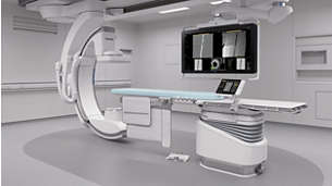
Azurion 7 M20 with FlexArm - the heart of our solution for cardiac care
Treat one more patient per day, by reducing procedure time by 17%* with optimized workflow options in interventional therapy and clinical software.

Live image guidance
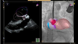
and fusion — EchoNavigator
Automatically fuses live 3D TEE and live X-ray in real time. So you can intuitively guide your device in the 3D space more quickly and efficiently when every move counts.

Insightful planning and guidance for SHD procedures - HeartNavigator
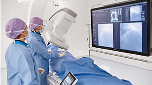
Stay close to the heart with increased confidence and ease during transcatheter aortic valve replacement (TAVR) and other challenging SHD procedures. The immersive user experience is highly automated to simplify planning, device selection and projection angle selection. During procedures, it provides live image guidance to support device positioning.
Technologies and innovations
-
![Clinically proven Azurion with ClarityIQ technology]()
Providing superb image quality at ultra-low dose settings.
Footnotes
*Reducing procedure time by 17%, with the ability to treat 1 more patient per day with optimized workflow options in image guided therapy and clinical software (Azurion - Philips Azurion Simulation Study 2016 - 12NC 452299123041 - FEB 2017). Results are specific to the institution where they were obtained and may not reflect the results achievable at other institutions. Please note that not all products displayed on our website may be available in your country due to varying local regulations and market conditions. For the most accurate availability information, please contact your local representative.


